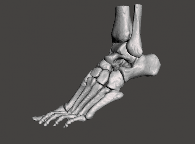
Human Left FootThe 3d model was created from computed tomography scans (CT medical data). It represents the human left foot.The feet are flexible structures of bones, joints, muscles, and soft tissues that let us stand upright and perform activities like walking, running, and jumping. They are divided into three sections:The forefoot contains the five toes (phalanges) and the five longer bones (metatarsals).The midfoot is a pyramid-like collection of bones that form the arches of the feet. These include the three cuneiform bones, the cuboid bone, and the navicular bone.The hindfoot forms the heel and ankle. The talus bone supports the leg bones (tibia and fibula), forming the ankle. The calcaneus (heel bone) is the largest bone in the foot.****************The model includes:- Tibia- Fibula- Talus- Calcaneus- Three cuneiform bones- Cuboid bone- Navicular bone- First, second, third, fourth, and fifth metatarsal bone- Phalanges(Please note that phalanges are not separated)****************Patient description: age 17, male.The model was created for educational use.Intends to help those interested in human anatomy and physiology during their learning process, as well as encourage other people deepen their knowledge about the human body.
Left Foot – male 3D Model